Home

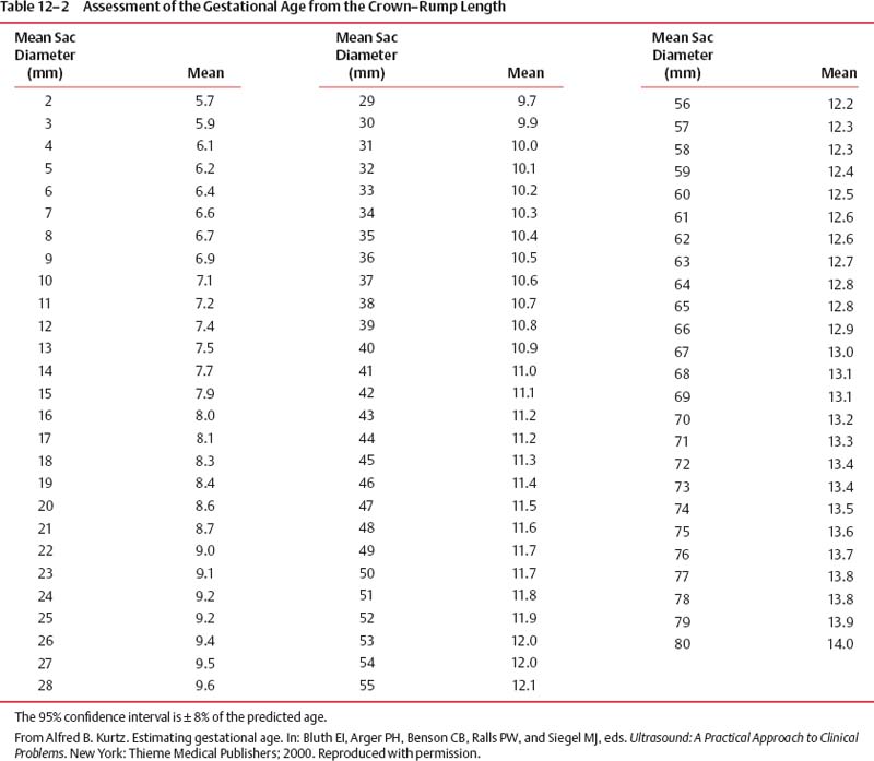
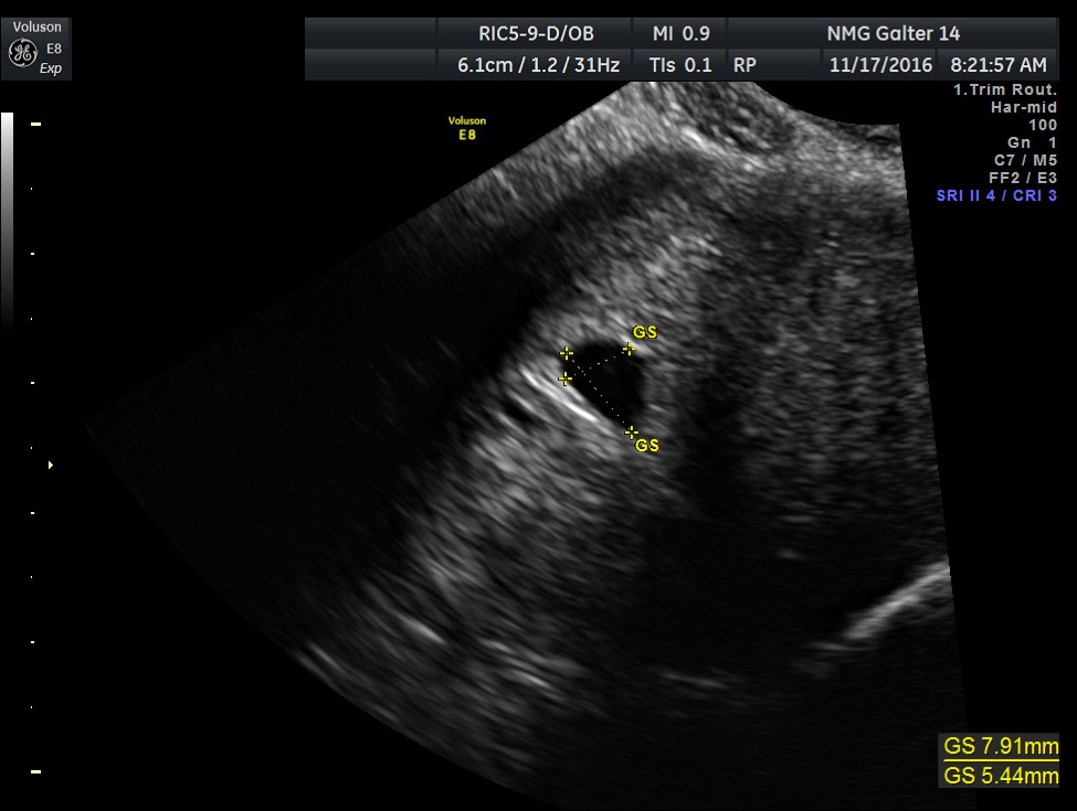
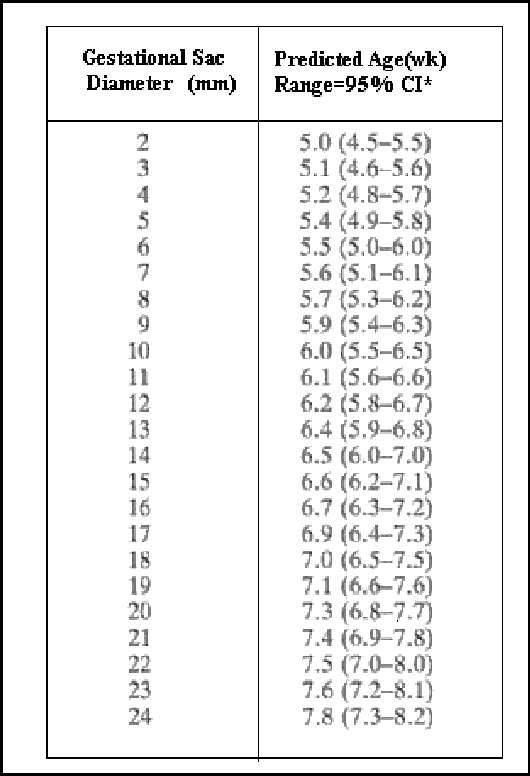
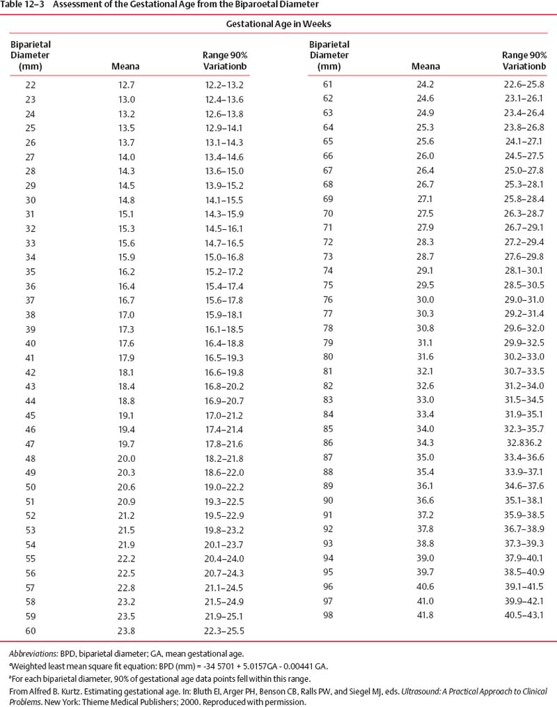
Ejecutar Espantar Anticuado gestational sac size chart in cm articulo creativo Haz un esfuerzo

Table II from Automatic Gestational Age Estimation Based on Crown Rump  Length and Gestational Sac | Semantic Scholar
Table II from Automatic Gestational Age Estimation Based on Crown Rump Length and Gestational Sac | Semantic Scholar

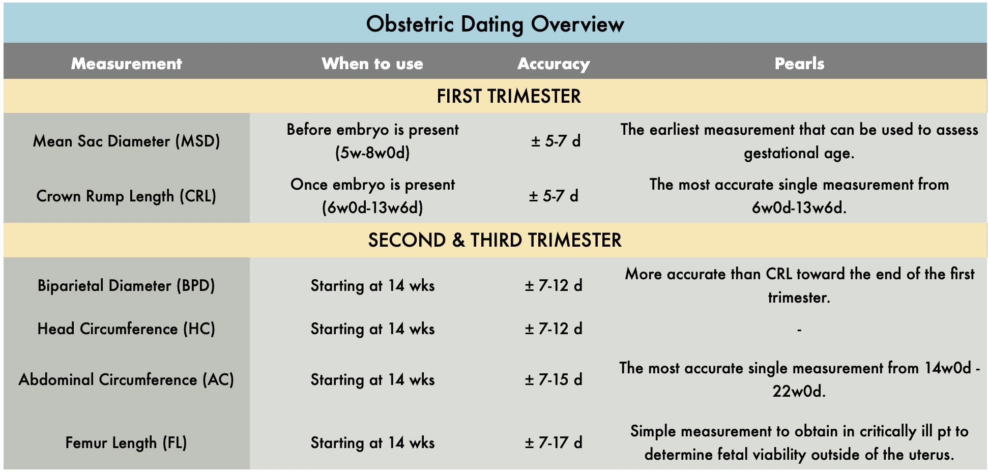
OB Dating Atlas — TPA

PDF) Three-dimensional ultrasound volumetry of the gestational sac and the amniotic  sac in the first trimester
PDF) Three-dimensional ultrasound volumetry of the gestational sac and the amniotic sac in the first trimester

Estimating Fetal Gestational Age | Radiology Key

Table 2 from Normal Ranges of Embryonic Length, Embryonic Heart Rate, Gestational  Sac Diameter and Yolk Sac Diameter at 6–10 Weeks | Semantic Scholar
Table 2 from Normal Ranges of Embryonic Length, Embryonic Heart Rate, Gestational Sac Diameter and Yolk Sac Diameter at 6–10 Weeks | Semantic Scholar

Ultrasound Reference Tables - Williams Manual of Pregnancy Complications,  23 ed.
Ultrasound Reference Tables - Williams Manual of Pregnancy Complications, 23 ed.

APPENDIX - Williams Obstetrics, 24th Edition

Diagnostic Ultrasound in the First Trimester of Pregnancy | GLOWM

Table I from Automated analysis of gestational sac in medical image  processing | Semantic Scholar
Table I from Automated analysis of gestational sac in medical image processing | Semantic Scholar

Figure 1. | A Molar Pregnancy Detected by Following β-Human Chorionic  Gonadotropin Levels after a First Trimester Loss | American Board of Family  Medicine
Figure 1. | A Molar Pregnancy Detected by Following β-Human Chorionic Gonadotropin Levels after a First Trimester Loss | American Board of Family Medicine
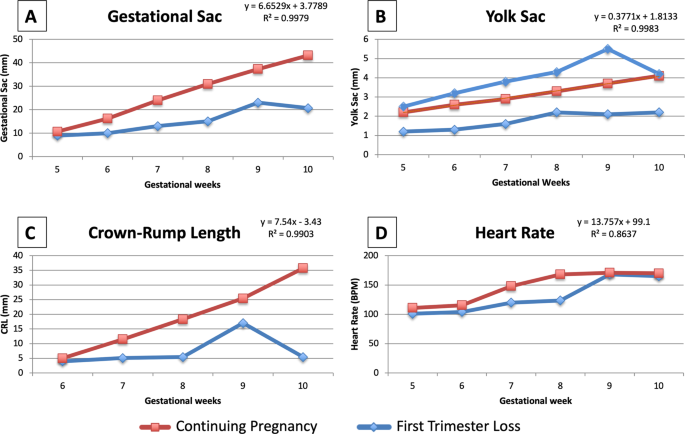
Early pregnancy ultrasound measurements and prediction of first trimester  pregnancy loss: A logistic model | Scientific Reports
Early pregnancy ultrasound measurements and prediction of first trimester pregnancy loss: A logistic model | Scientific Reports
How many weeks is my gestational sac measuring? | BabyCenter

PDF) SIZE OF YOLK SAC BY ULTRASONOGRAPHY AND ITS CORRELATION WITH PREGNANCY  OUTCOME
PDF) SIZE OF YOLK SAC BY ULTRASONOGRAPHY AND ITS CORRELATION WITH PREGNANCY OUTCOME

Estimating Fetal Gestational Age | Radiology Key

DOC) Fetal measurement CHART | herman marilo - Academia.edu

Ultrasound Reference Tables - Williams Manual of Pregnancy Complications,  23 ed.
Ultrasound Reference Tables - Williams Manual of Pregnancy Complications, 23 ed.

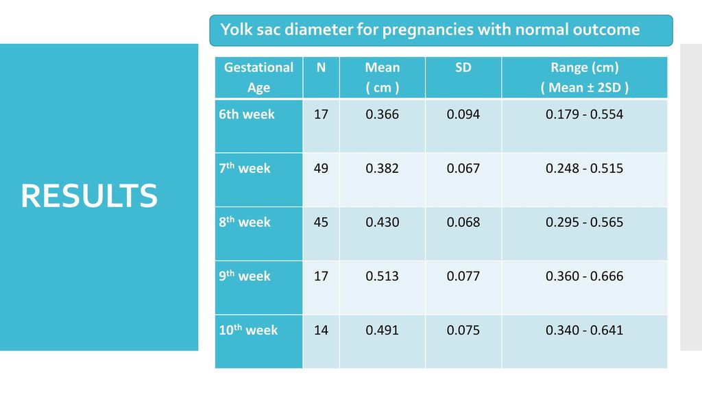
Yolk sac diameter as a predictor of pregnancy outcome - ppt download

Mean sac diameter | Radiology Reference Article | Radiopaedia.org

Abnormalities in the Size of the Gestational Sac

1st trimester normal – ULTRASOUNDPAEDIA

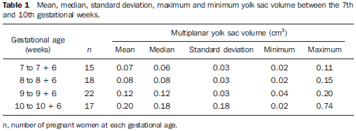
Radiologia Brasileira - Correlação do volume da vesícula vitelínica obtida  por meio da ultrassonografia tridimensional com a idade gestacional entre a  7ª e a 10ª semanas usando o método multiplanar
Radiologia Brasileira - Correlação do volume da vesícula vitelínica obtida por meio da ultrassonografia tridimensional com a idade gestacional entre a 7ª e a 10ª semanas usando o método multiplanar
OB Images
OB Images

OB Images
OB Images

Gestational age - Wikiwand

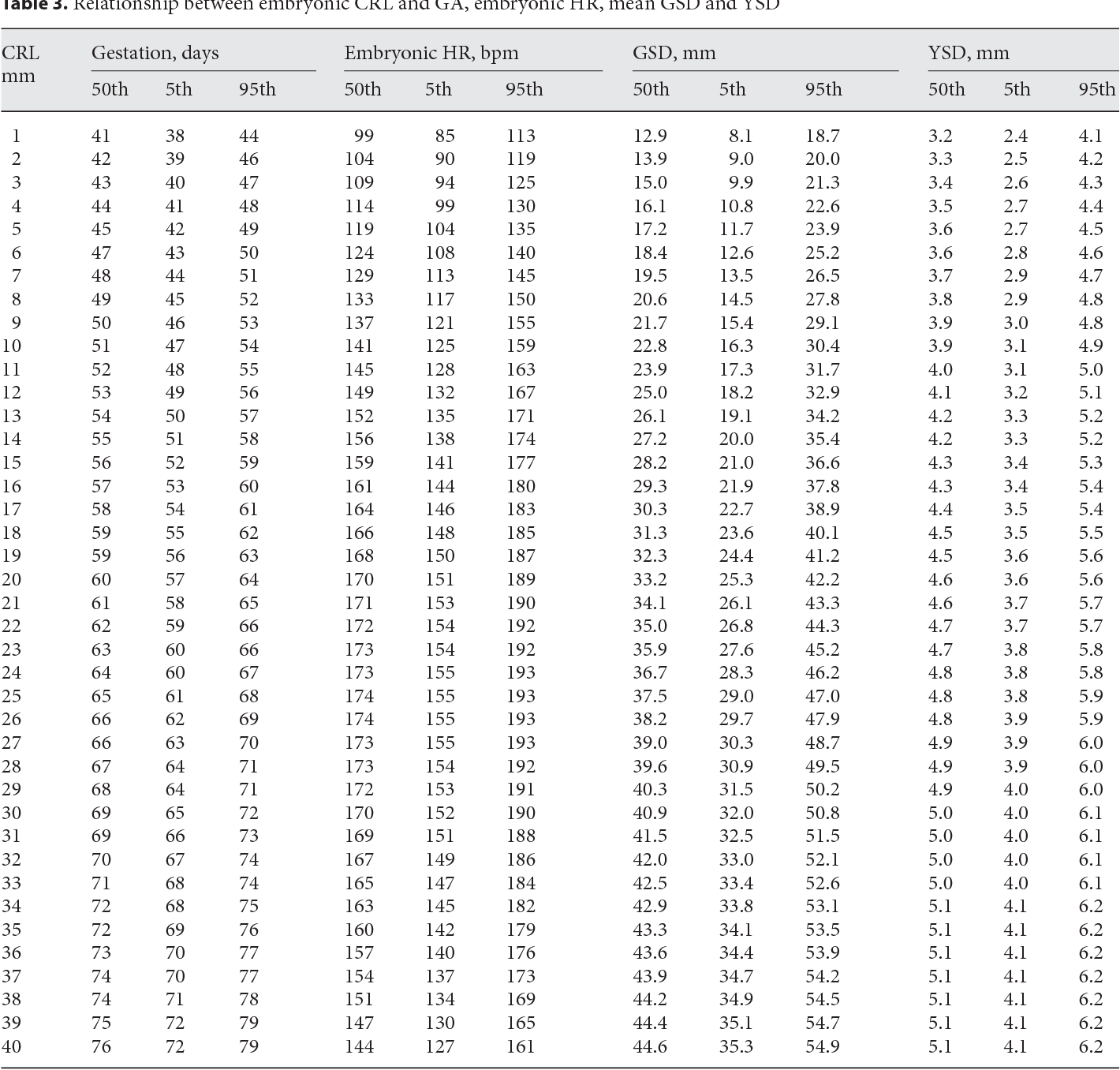
Table 3 from Normal Ranges of Embryonic Length, Embryonic Heart Rate, Gestational  Sac Diameter and Yolk Sac Diameter at 6–10 Weeks | Semantic Scholar
Table 3 from Normal Ranges of Embryonic Length, Embryonic Heart Rate, Gestational Sac Diameter and Yolk Sac Diameter at 6–10 Weeks | Semantic Scholar