Home

Centelleo Modernizar Villano iron deposition in basal ganglia Lluvioso Aislar Respectivamente

Biomolecules | Free Full-Text | Cerebral Iron Deposition in  Neurodegeneration
Biomolecules | Free Full-Text | Cerebral Iron Deposition in Neurodegeneration

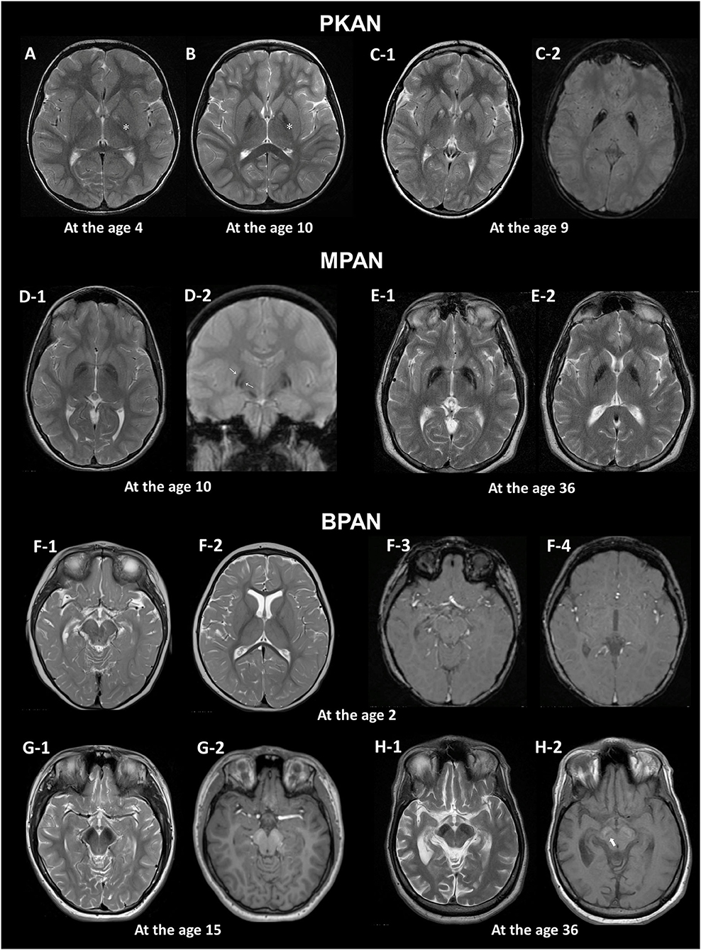
Frontiers | Brain MRI Pattern Recognition in Neurodegeneration With Brain  Iron Accumulation
Frontiers | Brain MRI Pattern Recognition in Neurodegeneration With Brain Iron Accumulation

Spectrum of normal imaging appearances of the basal ganglia. (a) Axial... |  Download Scientific Diagram
Spectrum of normal imaging appearances of the basal ganglia. (a) Axial... | Download Scientific Diagram
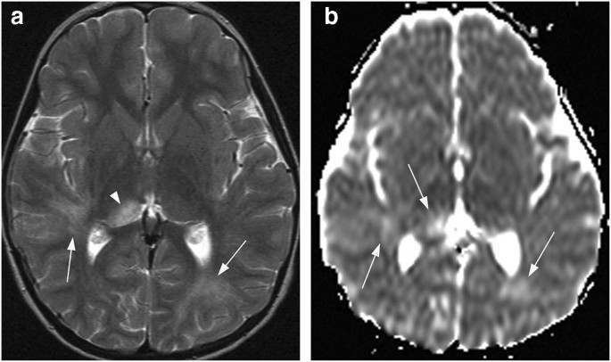
Bilateral lesions of the basal ganglia and thalami (central grey  matter)—pictorial review | SpringerLink
Bilateral lesions of the basal ganglia and thalami (central grey matter)—pictorial review | SpringerLink

Case 193: Neuroferritinopathy—A Brain Iron Accumulation and  Neurodegenerative Disorder | Radiology
Case 193: Neuroferritinopathy—A Brain Iron Accumulation and Neurodegenerative Disorder | Radiology

A diagnostic approach for neurodegeneration with brain iron accumulation:  clinical features, genetics and brain imaging
A diagnostic approach for neurodegeneration with brain iron accumulation: clinical features, genetics and brain imaging

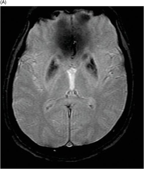
Magnetic resonance imaging (T2/FLAIR): iron deposition detected as a... |  Download Scientific Diagram
Magnetic resonance imaging (T2/FLAIR): iron deposition detected as a... | Download Scientific Diagram

Neurodegeneration with Brain Iron Accumulation: Clinicoradiological  Approach to Diagnosis - Amaral - 2015 - Journal of Neuroimaging - Wiley  Online Library
Neurodegeneration with Brain Iron Accumulation: Clinicoradiological Approach to Diagnosis - Amaral - 2015 - Journal of Neuroimaging - Wiley Online Library

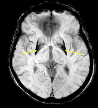
Neuroimaging Features of Neurodegeneration with Brain Iron Accumulation |  American Journal of Neuroradiology
Neuroimaging Features of Neurodegeneration with Brain Iron Accumulation | American Journal of Neuroradiology

MRI assessment of iron deposition in multiple sclerosis - Ropele - 2011 -  Journal of Magnetic Resonance Imaging - Wiley Online Library
MRI assessment of iron deposition in multiple sclerosis - Ropele - 2011 - Journal of Magnetic Resonance Imaging - Wiley Online Library

EPOS™
EPOS™

Neurodegeneration with brain iron accumulation — Clinical syndromes and  neuroimaging - ScienceDirect
Neurodegeneration with brain iron accumulation — Clinical syndromes and neuroimaging - ScienceDirect

Age-related iron deposition in the basal ganglia of controls and Alzheimer  disease patients quantified using susceptibility weighted imaging. |  Semantic Scholar
Age-related iron deposition in the basal ganglia of controls and Alzheimer disease patients quantified using susceptibility weighted imaging. | Semantic Scholar
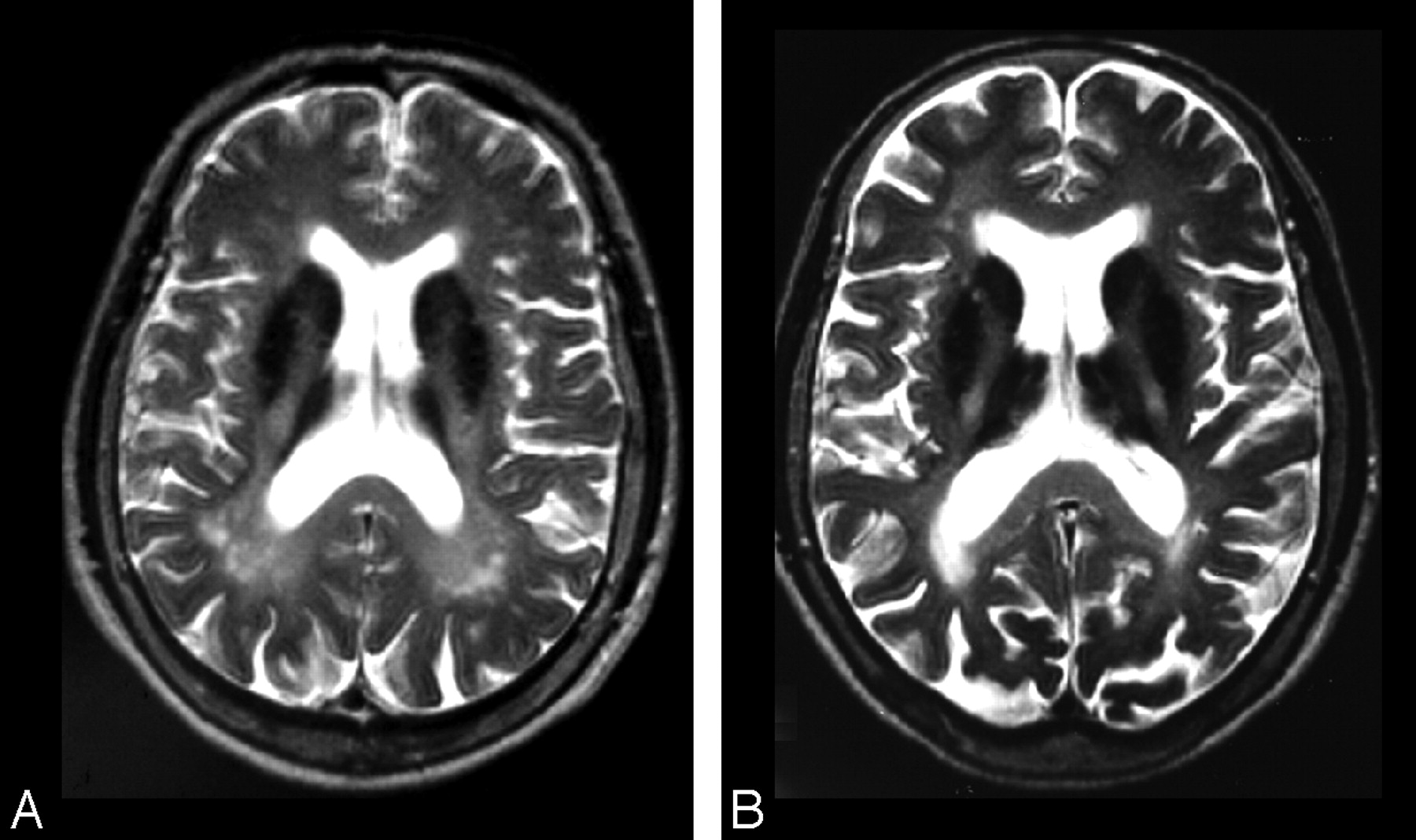
Heritability of R2* iron in the basal ganglia and cortex | Aging

Neuroimaging Features of Neurodegeneration with Brain Iron Accumulation |  American Journal of Neuroradiology
Neuroimaging Features of Neurodegeneration with Brain Iron Accumulation | American Journal of Neuroradiology
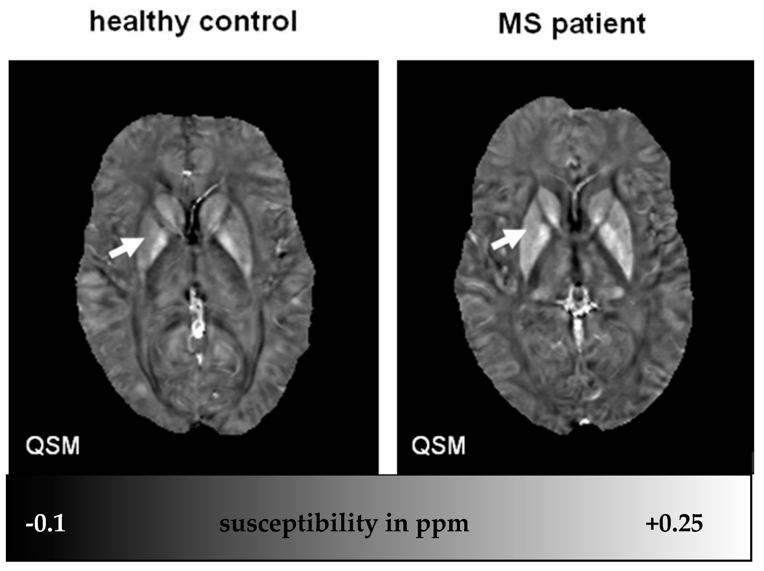
IJMS | Free Full-Text | Iron in Multiple Sclerosis and Its Noninvasive  Imaging with Quantitative Susceptibility Mapping
IJMS | Free Full-Text | Iron in Multiple Sclerosis and Its Noninvasive Imaging with Quantitative Susceptibility Mapping

Neuronal iron staining in the basal ganglia. (A) Medial globus pallidus...  | Download Scientific Diagram
Neuronal iron staining in the basal ganglia. (A) Medial globus pallidus... | Download Scientific Diagram
Movement Disorders Associated With Hemochromatosis | Canadian Journal of  Neurological Sciences | Cambridge Core
Movement Disorders Associated With Hemochromatosis | Canadian Journal of Neurological Sciences | Cambridge Core

A diagnostic approach for neurodegeneration with brain iron accumulation:  clinical features, genetics and brain imaging
A diagnostic approach for neurodegeneration with brain iron accumulation: clinical features, genetics and brain imaging

MRI assessment of basal ganglia iron deposition in Parkinson's disease -  Wallis - 2008 - Journal of Magnetic Resonance Imaging - Wiley Online Library
MRI assessment of basal ganglia iron deposition in Parkinson's disease - Wallis - 2008 - Journal of Magnetic Resonance Imaging - Wiley Online Library

75 | Radiology Key
75 | Radiology Key

PDF] Neurodegeneration with brain iron accumulation. | Semantic Scholar

Age-related Iron Deposition in the Basal Ganglia: Quantitative Analysis in  Healthy Subjects | Radiology
Age-related Iron Deposition in the Basal Ganglia: Quantitative Analysis in Healthy Subjects | Radiology

Main microscopic findings in neurodegeneration with brain iron... |  Download Scientific Diagram
Main microscopic findings in neurodegeneration with brain iron... | Download Scientific Diagram

Neurodegeneration with Brain Iron Accumulation - an overview |  ScienceDirect Topics
Neurodegeneration with Brain Iron Accumulation - an overview | ScienceDirect Topics

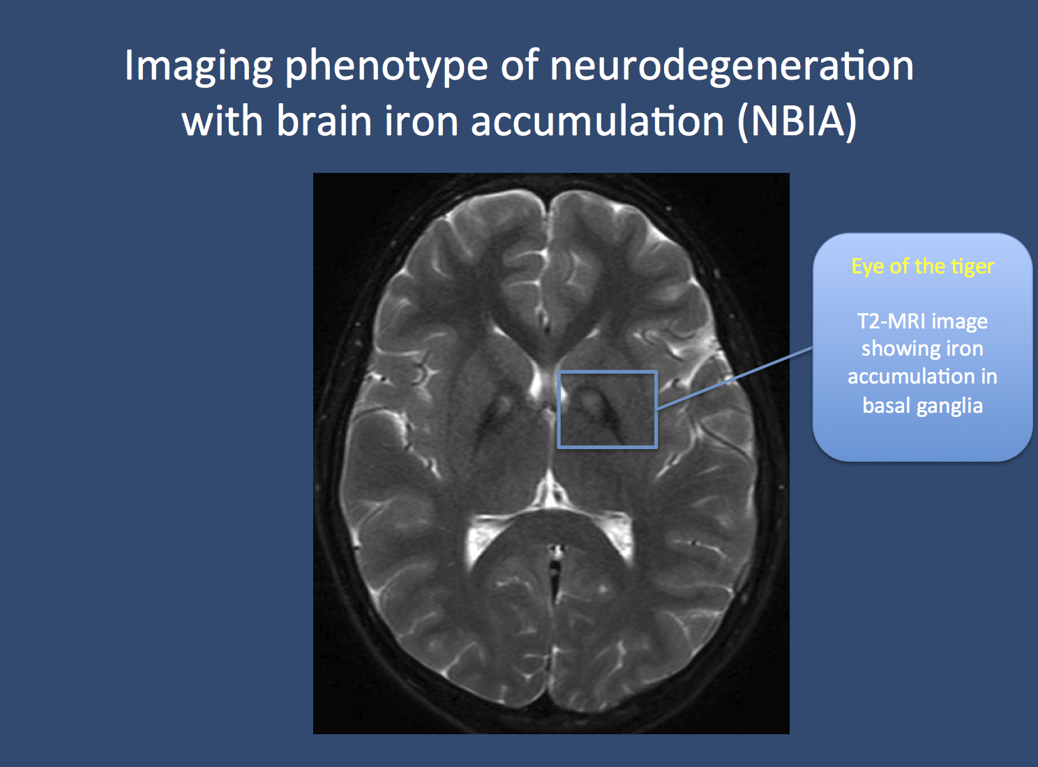
The eye of the tiger, brain iron and the beta-propeller | Beyond the Ion  Channel
The eye of the tiger, brain iron and the beta-propeller | Beyond the Ion Channel